
Figure 3.
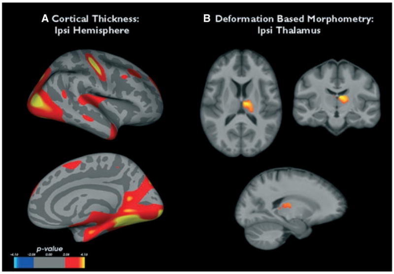
(A) Regions of cortical thinning in the ipsilateral hemisphere of temporal lobe epilepsy with mesiotemporal sclerosis (TLE-MTS) compared to the control group. (B) Thalamic regions showing significant volume loss in TLE-MTS compared to controls. See text for statistical thresholds.
Epilepsia © ILAE