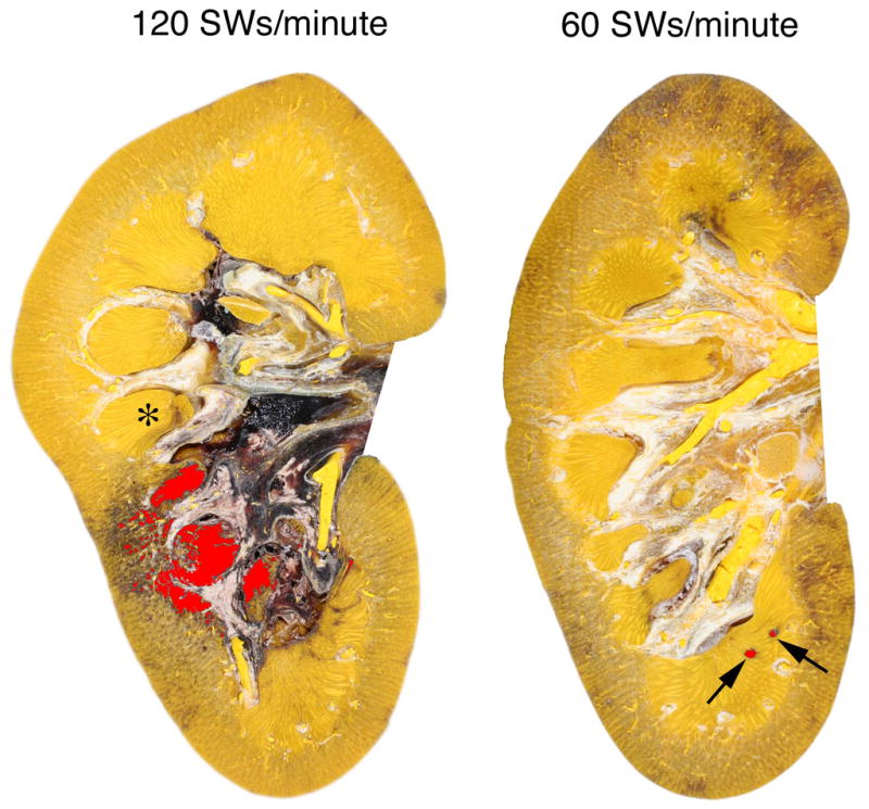
FIG. 1.

Representative cross-sections from pig kidneys treated at 120 or 60 SWs/min (2000 SWs, 24 kV) with an unmodified Dornier HM-3 lithotripter. The lesions have been coloured (red) to visualize regions of hemorrhage. Quantitation of the lesion was restricted to the renal parenchyma and did not include areas of bleeding into the renal pelvis, seen here as black deposits in the renal pelvicalyceal system. One papilla is marked with an asterisk (*). Damage to the kidney treated at 120 SWs/min involves several renal papillae and extends from the medulla to the cortex. The subcapsular hematoma in this kidney is beyond the plane of section. The lesion in the kidney treated at 60 SWs/min is restricted to small foci in the medulla (arrows).
