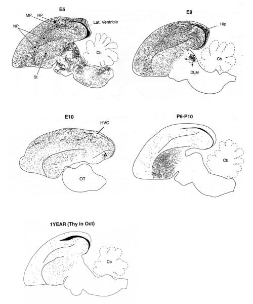
Figure 2.
The distribution of [3H]-labelled neurons in sagittal brain sections from 1-year old canaries following [3H]-thymidine injections at the embryonic (E) ages indicated. The lower map is from a 1-year old adult that received [3H]-thymidine injections in October and was then killed 125 days later. Although there is an overall decrease in neurogenesis (cell production + survival) as development proceeds, the absolute magnitude of the decrease cannot be ascertained from these figures due to several methodological factors, including variation in the amount of time [3H]-thymidine was available for cell labeling (see Alvarez-Buylla et al., 1994). However, developmental shifts in the regional distribution of new neurons are significant. Between E5 and E9, sub-telencephalic neuron addition is virtually over with the notable exception of DLM (medial portion of the dorsolateral nucleus of the thalamus), part of the vocal control system. Injections on E10 labelled cells restricted to the telencephalon, yet few such cells were found in HVC. As with DLM, HVC neurogenesis is delayed relative to surrounding regions. Post-hatching (P6-P10), large numbers of neurons are added to the striatum, including song system nucleus Area X (not shown). The most notable difference between birds injected as juveniles and adults is a greater decline in striatal, relative to extra-striatal, neuron addition in older birds. Nevertheless, neuron addition is widespread even in adults. Dorsal = up and Rostral = left, Modified from (Alvarez-Buylla et al., 1994).

