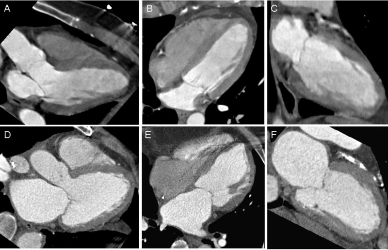
Figure 1.

Three-chamber (A), four-chamber (B) and two-chamber (C) views obtained at end-systole of a patient in the control group, without acute coronary syndrome, and in the lowest quartile of LAVmax. Corresponding views (D–F) in a patient in the “risk factor” group, with acute coronary syndrome, and in the top quartile of LAVmax. LAVmax denotes maximal left atrial volume.
