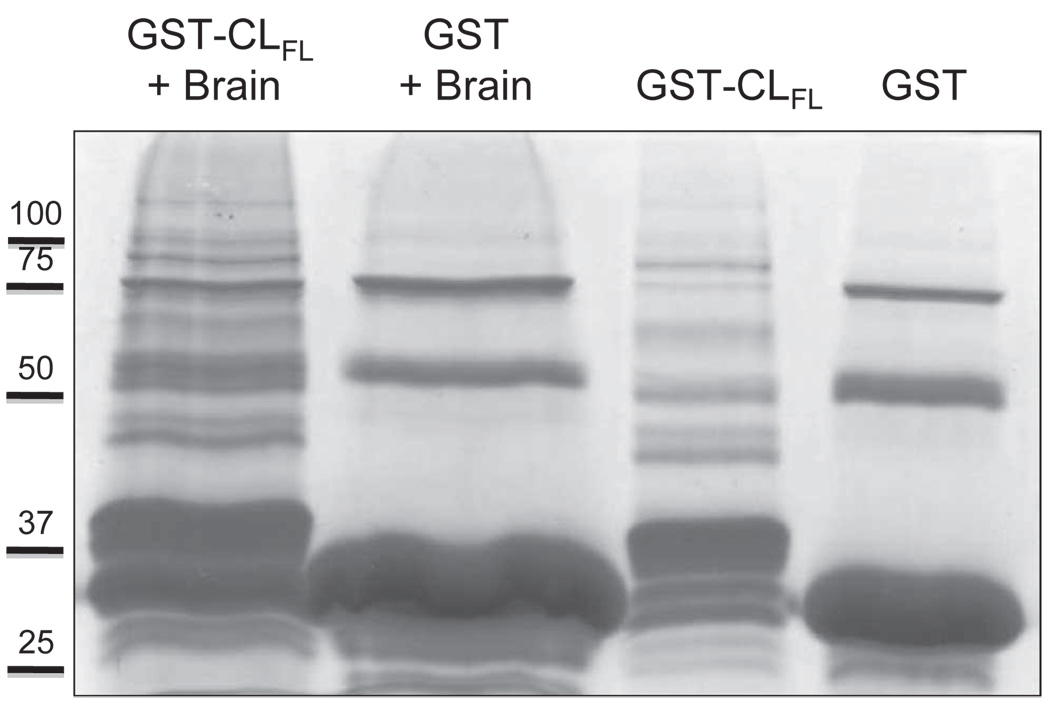
Figure 2.
Coomassie staining of protein samples obtained from glutathione-S-transferase (GST) pull-down assays. Products used in the assay included the full-length cytoplasmic loop (CLFL) sequence of Cx43 fused to GST and GST alone. These products were allowed to bind glutathione sepharose beads and were then eluted and collected (right-most two lanes) or exposed to mouse brain lysate prior to elution (left-most two lanes). Excess protein bands in the left-most lane containing eluate from the GST-CLFL beads exposed to brain lysate suggest novel proteomic interactions involving the Cx43-CLFL.

