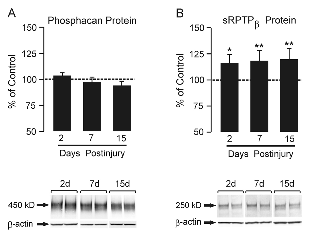
Figure 1. Time course of whole hippocampal phosphacan and RPTPβ protein expression at 2, 7 and 15d after UEC.
Western blot for phosphacan (A) within the deafferented hippocampus showed no difference from control at any of the three time points. 2d and 15d n=6; 7d n=9. In (B), Western blot probe for RPTPβ (antibody recognizing both full length and short transmembrane phosphatase) showed a significant increase in protein within the deafferented hippocampus at all time points when compared with contralateral controls. Representative ipsilateral (left)/contralateral (right) gel pairs are illustrated below each time point. 2d and 15d n=6; 7d n=9; *p<0.02, **p<0.001.

