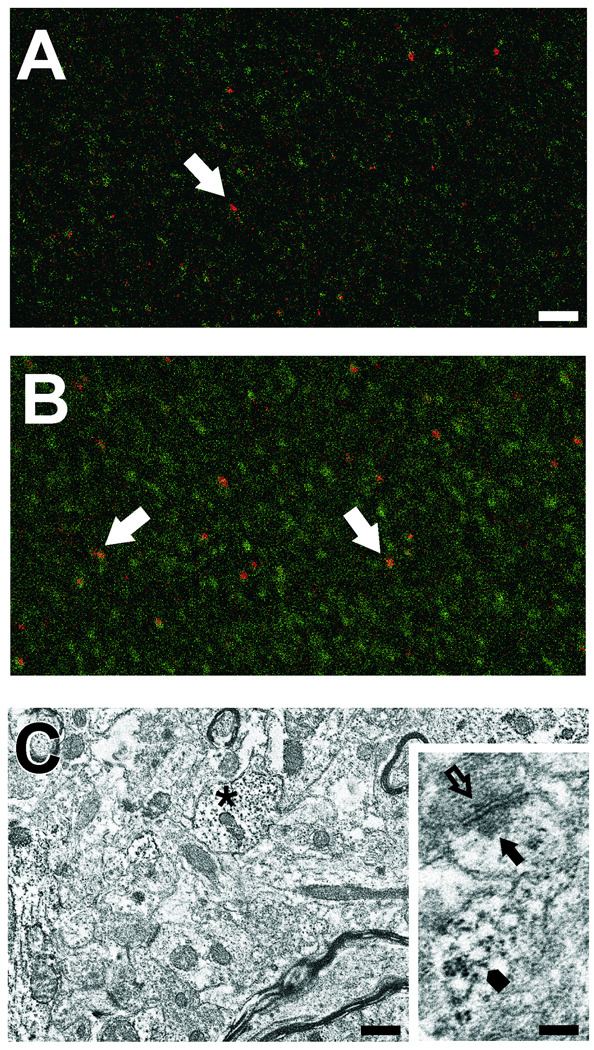
Figure 3. RPTPβ distribution in dentate ML at 7d after UEC lesion.
Confocal IHC shows RPTPβ (green) at punctate sites in the outer contralateral ML (A). These sites are more numerous and of larger size in the deafferented ML (B), where RPTPβ is found adjacent to postsynaptic sites (PSD-95 positive profiles, red; arrows in A,B). Ultrastructural ICC shows RPTPβ localized in dendritic profiles (asterisk) in the outer ML of a contralateral hemisphere (C). Inset in C shows RPTPβ present in a spine (arrowhead), postsynaptic (arrow) and presynaptic (open arrow) sites from a section without counter stain. Bar in A, B=25µm; C=0.5µm; inset=0.2µm.

