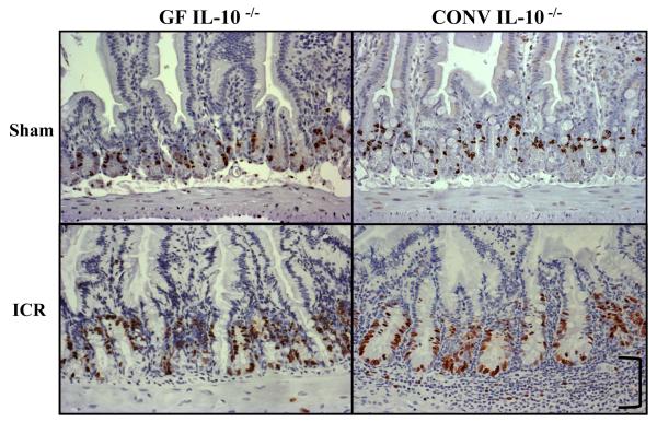
Figure 3. Operative BrdU Immunohistochemistry.
Representative BrdU immunostained sections from the jejunum of sham-operated and ICR mice 7d following operation (20x). Note the increases in BrdU labeled cells and crypt depth that occur in both GF and CONV mice after ICR. In addition, marked submucosal inflammation (bracket) is only present in the CONV mouse after ICR when compared to GF and sham-operated groups.

