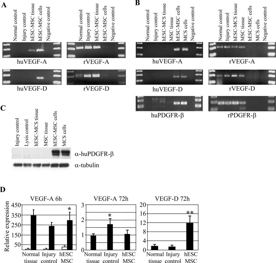
Figure 4.
(A) RT-PCR showing species-specific VEGF-A and VEGF-D amplification at 6-hour time point. Human specific primers show hESC-derived MSC-originated VEGF-A amplification in transplanted tissues and in positive cell controls. (B) RT-PCR analysis at 72-hour time point. VEGF-A, VEGF-D, and PDGFR-β amplification were seen only with rat specific primers in transplanted tissues. (C) Western blot analysis for human PDGFR-β for 72-hour tissue homogenates. Only control MSC cells yielded a positive staining. α-Tubulin was used to normalize the samples. (D) Quantitative RT-PCR shows that majority of VEGF-A at 6-hour time point was synthesized by rat specific primers. White bars represent human specific amplification and black bars rat specific amplification. Quantification of the VEGF-A and VEGF-D production at 72-hour time point with primers recognizing both rat and human sequences show increased VEGF-D expression in hESC-derived MSC animals as compared to injured control group.

