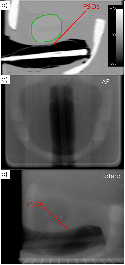
Fig. 7.

CT and kV images of the PSDs inserted into an anthropomorphic phantom. a) A sagittal CT slice with the prostate contoured (green), showing the PSDs. b) An anteroposterior kV image and c) a lateral kV image acquired with the linac’s OBI system.
