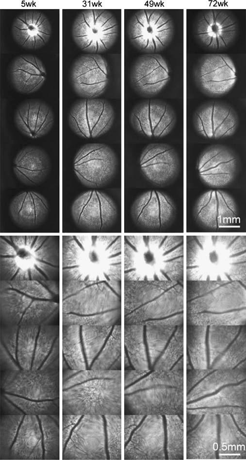
Fig. 2.
Long-term time-lapse fundus fluorescence imaging with a Thy1::CFP mouse in vivo. Fundus CFP fluorescence is imaged at indicated times, which are the age of the mouse. Some areas in an image are not in focus and appear blurry due to spherical errors and refractive errors in the optics which have not been corrected. This sequence is a representative of four separate mice which all show similar results. Bar, 500 μm.

