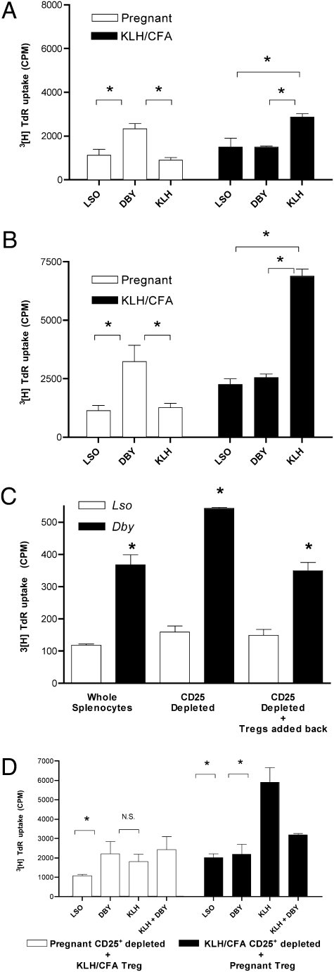
Fig. 4.
Pregnancy-induced T regulatory cells suppressive function is antigen-specific. (A) Splenocytes harvested from a timed first pregnancy of 6-week-old C57BL/6 × C57BL/6 male at dpc 18.5 (four male fetuses) as well as virgin 6-week-old C57BL/6 female immunized with KLH/CFA 7 days prior. Cellular proliferation assay was performed using 1 × 10⁶ cells per mL to the CD4+ T cell peptide epitope of the H-Y antigen presented by I-Ab (10 μM Dby), the I-Ab presented lysteriolysin peptide epitope (10 μM Lso) or KLH (1 μg/mL). Cultures were pulsed with 1 μCi per well 3[H]TdR for the final 18 h of a 72-h culture. (B) CD25+ reduced splenocytes. (C) Splenocytes harvested from a timed first pregnancy of 6 weeks – old C57BL/6 x C57BL/6 male at dpc 15.5 (three male fetuses). CD25+ cell reduced. CD25+ cells (Treg) added back to CD25+-depleted splenocytes in a ratio of 4:1. (D) CD25+-reduced splenocytes from either the dpc 18.5 mouse (□) or the KLH/CFA-immunized mouse (■) above cocultured with the CD25+ (Treg enriched population) isolated by magnetic bead positive selection from the KLH/CFA-immunized mouse (□) or the pregnant mouse (■) at a ratio of 4:1. Stimuli as indicated (Lso 10 μM, Dby 10 μM, KLH 1 μg/mL). Statistical difference determined by one-way ANOVA with Bonferroni–Dunn posttest (N.S., nonsignificant; *, P < 0.05).

