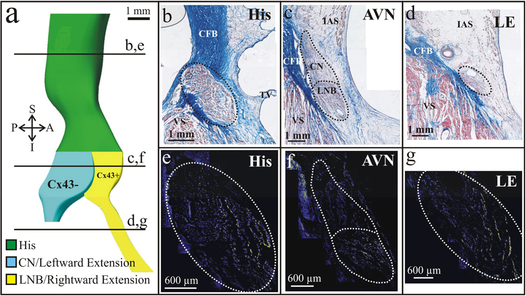
Figure 2. Cx43 expression in the Human AVJ.
Histological serial sections revealed the presence of rightward and leftward posterior nodal extensions. In addition, immunofluorescence identified differences in the expression of Cx43 between the two extensions and their corresponding counterparts in the AV node. Immunostaining of the His bundle also identified a gradient of Cx43 expression in line with the differential expressions of the protein in the CN and LNB in this particular preparation. (a) 3D reconstruction of an AVJ from a 58 year old female. (b–d) Histological sections as denoted in (a) by the solid black lines. (e–g) Corresponding immunostained sections for Cx43. Yellow color shows Cx43.

