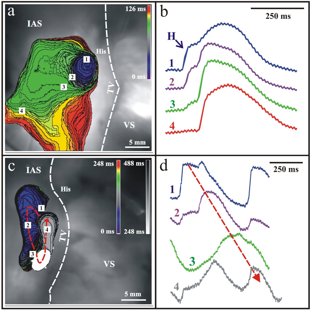
Figure 3. Junctional Rhythm and Reentry in the Human AVJ.
Optical mapping with voltage-sensitive dyes revealed the site of origin of the AVJ rhythm to be in the NH region. (a) The leading pacemaker, in this preparation, produced anterograde excitation towards His bundle (not shown) and retrograde excitation towards the compact node. Retrograde excitation spread in the compact AV node and excited the atrium via the fast pathway. (b) Corresponding optical action potentials to the observed junctional rhythm. (c) In another case, retrograde excitation spread over the Cx43-negative leftward nodal extension, turned around and reentered into the Cx43-positive rightward nodal extension. The excitation returned to the site of origin, completing one reentrant circuit. (d) Corresponding optical action potentials confirming the presence of one reentrant circuit as denoted by the dotted arrow.

