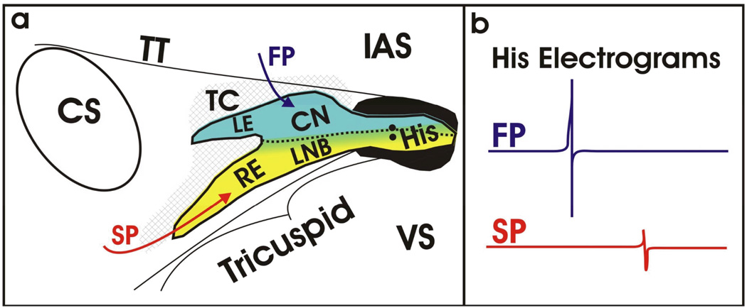
Figure 5. Structural and Functional Understanding of the AVJ.
Based on over a century of research, our current understanding of the AVJ is beginning to blend both function and structure as unified definition as shown in (a). The directional arrows illustrate the functional inputs to the AVJ (fast pathway (FP) in red, slow pathway (SP) in blue) roughly corresponding to the Cx43-positive and –negative regions as defined structurally. (b) Representative His electrograms resulting from activation of the fast and slow pathways, in blue and red, respectively. The significant difference in amplitude results from differential compartments within the AVJ being activated during conduction.

