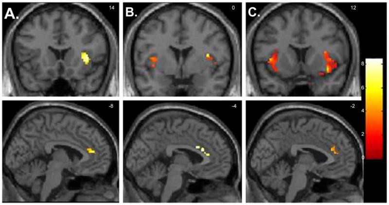
Fig. 1.

Selected regions of interest where there were foci of significantly decreased neural activity when healthy volunteers (n=12) were medicated versus unmedicated while viewing emotion-provoking paradigm (paired t-tests, p<0.05 corrected for multiple comparisons). Foci of significance were overlaid on coronal and sagittal brain slices spatially normalized into the Montreal Neurologic Institute coordinates system. A. Happiness paradigm; B. Fear paradigm and C. Anger paradigm. Significant differences were detected across the three emotion-provoking paradigms in the insula (shown on the sections on the superior side of the illustration), and in the anterior cingulate gyrus (shown on the inferior slices). Brain slices correspond to coronal view (superior panel) an sagittal view (inferior panel). Left side of the brain corresponds to the left side of the picture. The numbers at the right upper corner at each frame represent standard coordinates in the y (coronal view) and x (sagittal view) axis.
