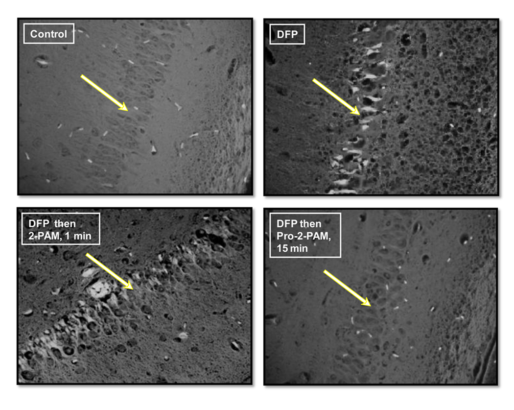
Fig. 5.
Fluoro-jade staining of representative guinea pig brains (40× magnification) for the hippocampus pyramidal neuron layer (lower CA1–CA2 region) at 24 h. White arrows point to the hippocampus neuron layer. Apoptotic cells, highlighted by fluorescence above background, are distinctly seen in the DFP and DFP then 2-PAM treatment 1 min after OP exposure.

