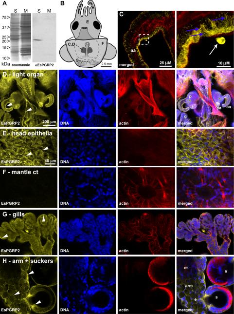
Figure 2. The localization of EsPGRP2 in E. scolopes tissues.
(A) The anti-EsPGRP2 antibody reacts with a single major protein species in the total soluble fraction of extracts from whole juvenile E. scolopes. The estimated molecular mass of the band was 20.5 kDa, consistent with the predicted mass of 20.8 kDa for the mature form of EsPGRP2. S, soluble fraction; M, membrane fraction. (B) Multiple tissues of juvenile E. scolopes were examined for expression of the EsPGRP2 protein by confocal immunocytochemistry, including the anterior appendage (C) of the light organ (D), the head epithelia (E), connective tissue of the mantle, including a chromatophore (F), the gills (G), and the arm and suckers (H). Juvenile E. scolopes were probed with the anti-EsPGRP2 antibody (yellow), and counterstained for actin-cytoskeleton (rhodamine-phalloidin) and DNA (TOTO-3). The EsPGRP2-antibody stained the apical portion of epithelial cells with the greatest intensity (arrowheads) and the staining appeared granular (blue arrows). Dashed box in left panel of C indicates area of the light-organ anterior appendage shown in higher magnification in right panel. Occasional extracellular protein staining was observed (white arrow). aa, anterior appendage; ct, connective tissue; e, epithelial cells of the arm; g, gills; hg, hindgut; p, pores; pa, posterior appendage; s, suckers.

