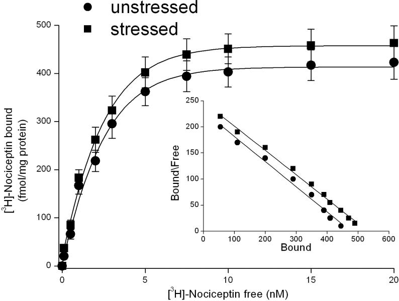
Figure 2.
Saturation curves and Scatchard plot of [3H]-Nociceptin binding in dorsal raphe nucleus slices from unstressed rats and from rats submitted to 15 min of forced swim (stressed rats). Saturation binding experiments were performed as described in Materials and Methods. The points represent the means ± s.e.m. of 5 experiments.

