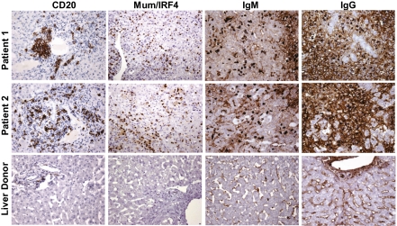
Fig. 3.
Immunohistochemical staining of B cell lineage markers in liver tissue from two patients with HBV-associated ALF and a representative control liver donor. Liver specimens obtained at the time of OLT from the native livers of patient 1 with massive hepatic necrosis and patient 2 with submassive hepatic necrosis, and from a control liver donor, were stained with monoclonal antibodies against CD20, MUM1/IRF4, IgM, and IgG with the use of immunoperoxidase. Sections from patient 1 and patient 2 show the presence of B cell clusters (CD20) around the portal areas; strong nuclear staining for MUM1/IRF4 is seen in a large number of plasmacytoid cells and plasma cells diffusely distributed in the lobules; IgM-secreting mature plasma cells are seen predominantly in the lobules; IgG-secreting plasma cells show a greater range of differentiation with plasmacytoid cells containing more open chromatin and often a prominent nucleolus (Fig. S5 A and B). In addition, a diffuse staining for IgG, and to a lesser extent IgM, is seen in the intercellular spaces and in patient 2 on the surface of residual hepatocytes. Sections from the control liver show few CD20-positive cells within the portal space and limited Ig deposits in the sinusoids. (Magnification: ×75.)

