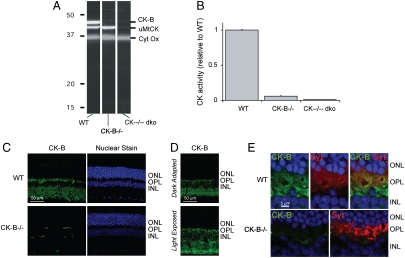
Fig. 1.
Identification of the types of CK in mouse retina. (A) Immunoblot of retina homogenates from WT, CK-B-/-, and CK--/-- dko mice. The blots were probed with antibodies to CK-B and uMtCK. The pattern of labeling demonstrates the specificity of the antibodies. (B) CK activity in retinal homogenates from WT, CK-B-/-, and CK--/-- dko mice. The absence of CK activity in the dko retinas shows that no other active CK isoforms of CK are expressed at significant levels in retina. (C) CK-B antibody immunostaining (Top) in WT (Top) and in CK-B-/- knockout (Bottom). There is intense labeling is in the photoreceptor synapse layer. The signals in the CK-B-/- retina come from the secondary antibody reacting with an unknown antigen in the retinal capillaries. The panels on the right show nuclear staining to demonstrate that there is no retinal degeneration in CK-B-/- retinas. (D) Comparison of the distribution of CK-B in light- vs. dark-adapted mouse retinas. (E) Pre- and postsynaptic localization of CK-B in the photoreceptor terminal layer. Sections were double labeled with antibodies to CK-B (Green) and to a presynaptic marker, synaptotagmin (Red). The top three panels show the outer nuclear layer (ONL), outer plexiform layer (OPL), and inner nuclear layer (INL) of a WT mouse retina. The bottom two panels show labeling of a CK-B-/- retina.

