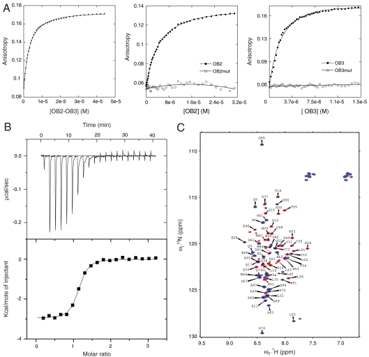
Fig. 2.
Binding of OB2, OB3, and OB2 + OB3 to p53 TAD. (A) Alexa fluor 546-labeled p53 TAD was titrated with OB2 + OB3, OB2, and OB3 in separate experiments. The data were fitted to a simple one-state binding model. (B) Binding isotherm of full-length p53 with OB3 as measured by using ITC in a buffer containing 25 mM Hepes, 150 mM NaCl, 3 mM DTT, pH 7.4 at 293 K. (C) Overlay of 1H15N HSQC of labeled p53 TAD (1–93) in the presence (Blue) and absence (Red) of OB3.

