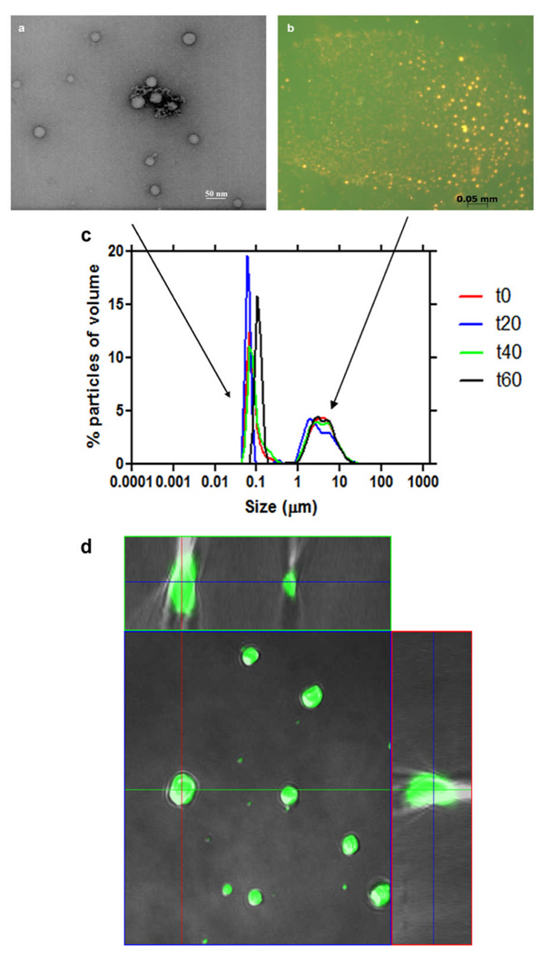
Fig. 1.
Characterization of monopalmitoyl glycerol in acetone/methanol solution after deposition onto an aqueous surface. Samples were extracted from just beneath the surface. TEM images show lipid particles in the nanometre size range (a), while fluorescence imaging of samples stained with the lipophilic stain, Nile Red (orange), and the water soluble dye, fluorescein (green), show particles in the micrometer size range (b). Measurements of lipid particle size distribution reveal two populations, one in the range 50 – 200 nm and the other 1 – 10 µm. This size distribution remained stable over a period of an hour (c). Confocal orthogonal projection confirms that lipid particles are not hollow, but rather continuous throughout their interiors (d).

