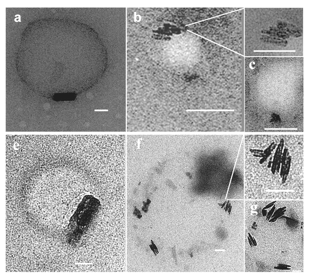
Fig. 6.
TEM of β-haematin formed at the MSG/water (a – c) and MPG/water (e – g) interface. A lipid and haematin solution was extruded through a fixed membrane filter and then introduced into an aqueous solution containing 50 mM citric acid (pH 4.8, 37 °C). Samples were taken for TEM images after 10 min of incubation. β-haematin crystals were observed aligned along the lipid-water interface for both extruded samples of MSG (top row) and MPG (bottom row). Clusters of β-haematin crystals were oriented parallel to each other and to the lipid-water interface (MSG, b, c; MPG, f, g).

