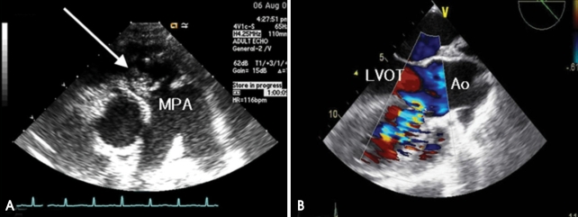
Fig. 1.
A: Transthoracic parasternal short axis view showing large, mobile vegetation (white arrow) on pulmonary valve. B: Transesophageal echocardiography showing a small subarterial type VSD with left to right shunt. Large amount of thrombus and vegetation were found in right ventricle. MPA: main pulmonary artery, LVOT: left ventricular outflow tract, Ao: aorta.

