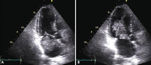
Fig. 1.
The echocardiogram performed at admission shows a villous left atrial myxoma that prolapses to the left ventricle in the diastolic phase. There is also akinesis of the mid to apical anterior walls, suggesting ischemia of the left anterior descending coronary artery. A: End-systolic phase. B: Middiastolic phase.

