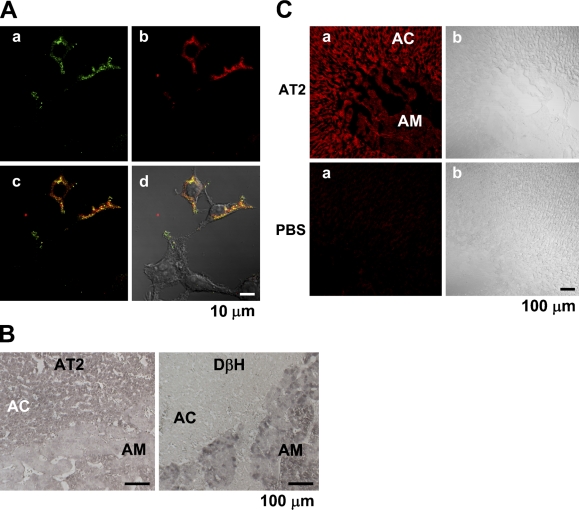
Figure 4.
Immunohistochemistry of bovine and rat adrenal glands for AT2. (A) Double staining for AT2 and hemagglutinin (HA) in HEK293T cells transfected with HA-AT2 vector. HEK293T cells were treated with rabbit AD-made anti-AT2 Ab (dilution, 1:50) and mouse anti-HA mAb (1:100) (A00186; GenScript, Piscataway, NJ). The reactions with the former and the latter were visualized with anti-rabbit IgG conjugated with Alexa 488 and anti-mouse IgG with Alexa 546, respectively. Panels a and b represent FITC-like and rhodamine-like fluorescence. Panel c is an overlay image of a and b with convergence in yellow, and d is an overlay image of c and differential interference contrast (DIC) image. (B) Immunostaining of bovine adrenal section with AD-made anti-AT2 Ab (1:100) and anti-DβH Ab (1:250) (AB1538; Chemicon). Each immunoreaction was visualized with the indirect immunoperoxidase method (see Materials and Methods). (C) Immunostaining of a rat adrenal section for AT2. The sections of rat adrenal gland were treated with or without (PBS) SC-made anti-AT2 Ab, and then with anti-rabbit IgG conjugated with Alexa 546.

