Figure 4.
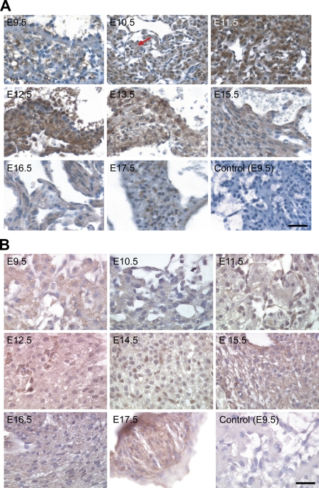
NcoA4 protein distribution in cardiac tissue during mouse development. Tissue distribution of NcoA4 protein in paraffin-embedded mouse embryo sections was determined by immunohistochemistry using rabbit anti-NcoA4 H300 (A) or goat anti-NcoA4 Q19 (B) polyclonal antibodies. All sections were counterstained with hematoxylin. NcoA4 immunoreactivity obtained with H300 was detected in ventricular cardiomyocytes (red arrow), with greater intensity observed from E11.5 to E13.5 (A). A similar but less-intense staining pattern was observed in cardiac tissue with the Q19 antibody (B). Absence of primary antibody or nonspecific IgG was used as a negative control, as shown for E9.5. Bars: A = 40 μm; B = 25 μm.

