Figure 5.
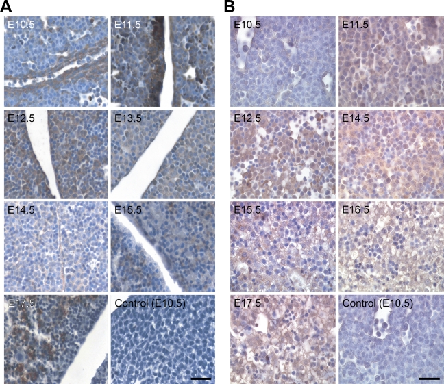
NcoA4 protein distribution in hepatic tissue during mouse development. Tissue distribution of NcoA4 protein in paraffin-embedded mouse embryo sections was determined by immunohistochemistry using rabbit anti-NcoA4 H300 (A) or goat anti-NcoA4 Q19 (B) polyclonal antibodies. All sections were counterstained with hematoxylin. The two compartments shown in A are two lobes of the liver. NcoA4 protein was expressed with high intensity in the hepatic tissue of the mouse embryo at E12.5 and E17.5, and less-intense NcoA4 staining was detected at E10.5 and E11.5 and from E13.5 to E15.5. Absence of primary antibody or nonspecific IgG was used as a negative control, as shown for E10.5. Bars: A = 40 μm; B = 25 μm.

