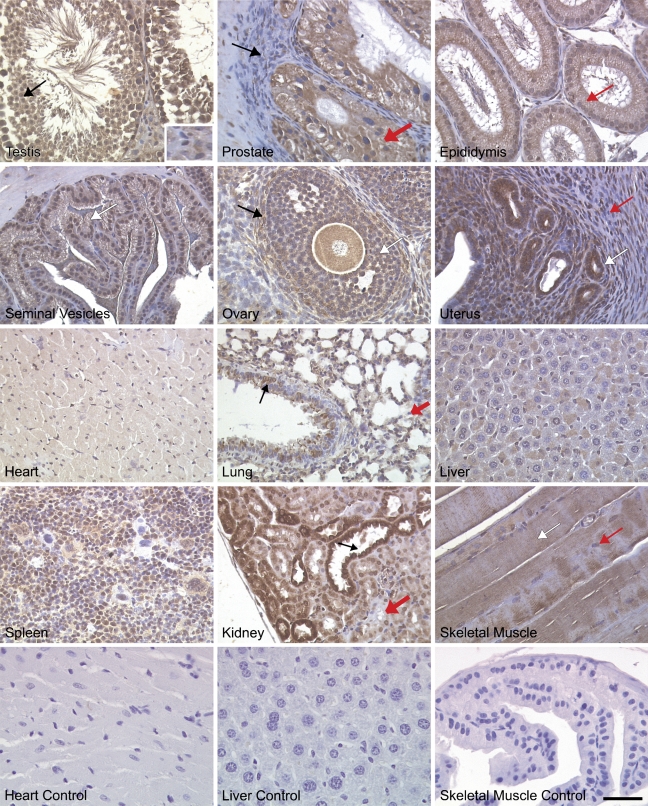
Figure 8.
NcoA4 protein distribution in various adult mouse tissues. Tissue distribution of NcoA4 protein on paraffin-embedded adult mouse tissue sections was determined by immunohistochemistry using anti-NcoA4 rabbit H300 polyclonal antibody. All sections were counterstained with hematoxylin. NcoA4 protein was highly expressed in testicular Sertoli cells (black arrow) and at all stages of spermatogenesis, whereas little staining was observed in Leydig cells (inset). In the prostate gland, glandular epithelial cells were stained positive, (thick red arrow), whereas staining was absent in the surrounding stroma (black arrow). Intense NcoA4 protein staining was detected in the columnar epithelial cells of the epididymis (red arrow). The columnar epithelial cells of the seminal vesicles were stained positive for NcoA4 (white arrow), whereas staining was absent in the muscular wall surrounding the lumen. NcoA4 protein was highly expressed in oocytes, granulosa cells (white arrow), and thecal cells surrounding the follicle (black arrow). However, the surrounding ovarian stroma was negative for NcoA4. Intense NcoA4 staining was detected in the glands of the uterus (white arrow), whereas little or no staining was detected in the circular (red arrow) smooth-muscle layer. NcoA4 protein was expressed in cardiac muscle but with less intensity than in the heart of the developing embryo. In the adult lung, both the bronchioles (black arrow) and alveoli (red thick arrow) stained positive for NcoA4 expression. Weak staining was detected in the liver, and strong staining was observed in the spleen. In the kidney, an intense staining for NcoA4 was detected in the distal ducts (black arrow) with less-intense staining in the proximal ducts (thick red arrow). Skeletal muscle fibers were positive (white arrow) for NcoA4, whereas the nuclei at the periphery of the fibers (red arrow) were negative. Staining in the absence of primary antibody was used as negative control for all tissues examined (not shown). Bar = 40 μm.

