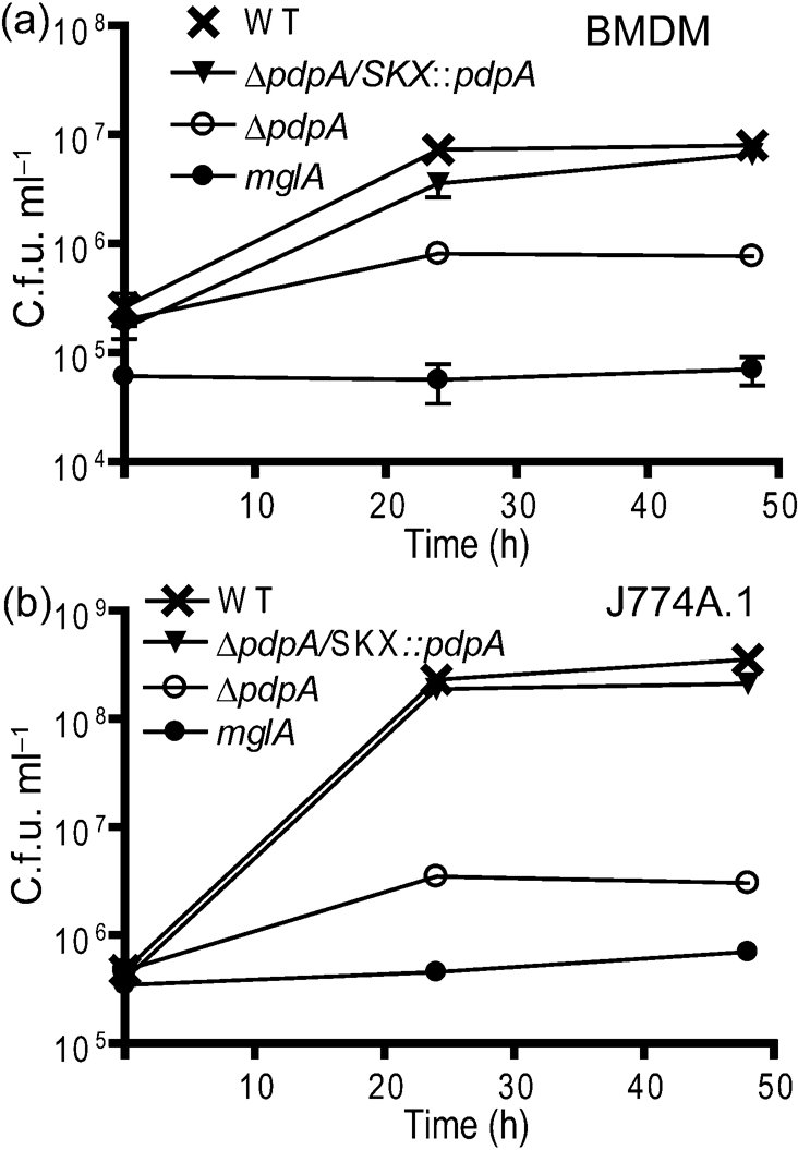
Fig. 1.

Intracellular growth of ΔpdpA mutants. The ΔpdpA mutant was able to replicate in BMDM (a) and J774A.1 cells (b) within the first 24 h of infection. Bacterial numbers decreased after 48 h and macrophages exhibited no signs of cytotoxicity. Complementation of the ΔpdpA deletion restored the wild-type (WT) growth phenotype. The ΔmglA mutant was included as a control as it is unable to replicate within macrophages. All data points in both panels are representative of three replicates and each experiment was performed in triplicate. Two-way ANOVA was used to calculate the significance of the differences in the growth curves between pairs of strains. For BMDM: WT vs ΔpdpA, P<0.00l; WT vs ΔpdpA/SKX : : pdpA, P=0.0261; and for ΔpdpA vs ΔpdpA/SKX : : pdpA, P=0.0028. For J774A.1 macrophages: WT vs ΔpdpA, P=<0.001; WT vs ΔpdpA/SKX : : pdpA, P=0.006; and for ΔpdpA vs ΔpdpA/SKX : : pdpA, P=<0.001.
