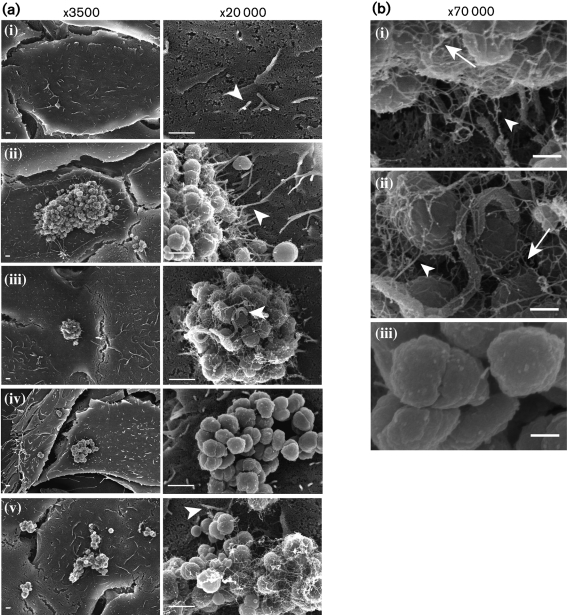
Fig. 1.
SEM imaging of human epithelial cells infected with N. gonorrhoeae. (a) A431 cells were mock-infected with medium (i), or infected with wt MS11(ii and iii), MS11pilE (iv) or MS11pilT (v) for 3 h. In each row, images in the two magnifications are from the same field of view. Arrowheads highlight microvilli shape/architecture as described in the text. Bars, 1 μm. (b) Enlarged images of wt MS11 (i and ii) and MS11pilE (iii) infected cells from (a). White arrows indicate Tfp connecting bacteria with each other. White arrowheads point to Tfp connecting bacteria to microvilli. Bars, 250 nm.

