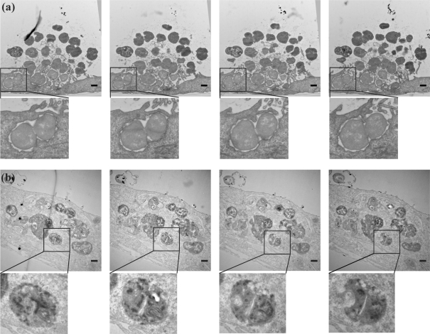
Fig. 4.
Serial thin-section imaging by TEM of extracellular and intracellular N. gonorrhoeae. A431 cells were infected with wt MS11 for 3 h (a) or 18 h (b). Successive serial thin-section (70 nm) images from each sample set are shown from left to right. Boxed regions in each image are enlarged below. Bars, 500 nm.

