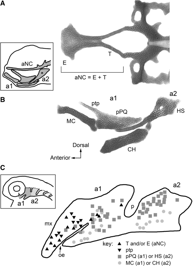
Fig. 3.
Fate map analysis of early larval zebrafish cartilages derived from the first and second pharyngeal arches. (A) Cartilages of the anterior neurocranium. Dissected preparation at 6 days postfertilization, in dorsal view. The boxed inset shows the position of the anterior neurocranium and pharyngeal cartilages in a left side view of the head. (B) Jaw (arch 1) and jaw supporting (arch 2) pharyngeal arch cartilages. (C) Fate map locations of the elements in the pharyngeal arches in the embryo at one day postfertilzation. The boxed inset shows the location of the arches in head, ventral, and posterior to the eye. The symbols in (C) represent individual cells, occasionally small groups of cells marked with a vital dye by electroporation and resulting in labeling of the cartilage elements as indicated in the key. a1, a2, arch 1 and 2; aNC, anterior neurocranium; CH, ceratohyal cartilage; E, ethmoid plate; HS, hyosymplectic cartilage; MC, Meckel's cartilage; mx, condensed crest of the maxillary region of a1; oe, oral ectodermal invagination (stomodeum); p, endodermal pharyngeal pouch 1 (hyomandibular pouch); pPQ, posterior region of the palatoquadrate; ptp, pterygoid process of the palatoquadrate; T, trabecula. Experiments by Mary Swartz, from Crump et al. (2004b, 2006) and Eberhart et al. (2006).

