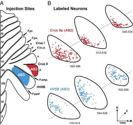
Fig. 2.
Injection sites and second-order neurons labeled in STN. (A) The injection sites of rabies virus (RV) with cholera toxin subunit β (CTb) are outlined on a flattened map of the cerebellar cortex adapted from ref. 7. The injection in AB2 (red filled area) targeted Crus IIp. The injection site in another animal (AB1, not illustrated) also targeted Crus IIp. In this case the injection site overlapped, but was somewhat less extensive than that of AB2. The injection in AB3 (blue filled area) targeted HVIIB. (B) Cross-sections of the STN show the location of second-order neurons labeled by the retrograde transneuronal transport of RV from Crus IIp in AB2 (red dots) and from HVIIB in AB3 (blue dots). Each of the three rostrocaudal levels displayed is spaced ≈1 mm apart. Labeled neurons from three consecutive sections (spaced 100 μm apart) are overlapped at each level. a, anterior; C, caudal; D, dorsal; F.amp., ansoparamedian fissure; F.in.cr., intracrural fissure; F.ppd., prepyramidal fissure; F.pr., primary fissure; F.ps., posterior superior fissure; M, medial; p, posterior.

