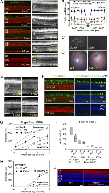
Fig. 2.
Bright light induces severe retinal degeneration in Palm−/− mice. (A) Palmitoylation-deficient (Palm−/−) and WT mice at 6 weeks of age were exposed to 10,000 lx light for periods indicated and then kept in the dark for 7 d, at which time SD-OCT and histological examinations were performed. Severe retinal degeneration was observed in Palm−/− mice whereas no degeneration was detected in WT retinas exposed to bright light for 60 min. INL, inner nuclear layer. (Scale bars: 20 μm.) (B) The thickness of the outer nuclear layer was measured 7 d after illumination with 10,000 lx for the indicated periods. ONH, optic nerve head. Error bars indicate SD of the means (n > 3). (C) Shown are representative outer retinal images obtained by SLO from 6-week-old Palm−/− mice exposed to 10,000 lx light for 5 min and then kept in the dark until examined 7 d later (Left) or from unexposed control mice kept in the dark (Right). Numerous autofluorescent retinal deposits are observed in illuminated Palm−/− mice. (D) Representative fundus images are shown from 6-week-old WT and Palm−/− mice taken 7 d after retinal illumination with 10,000 lx light for 60 min. Palm−/− mice manifest widespread atrophic changes. (E) SD-OCT B-scan imaging of the same eye performed at 1, 3, and 7 d after retinal exposure of 6-week-old Palm−/− mice to 10,000 lx light for 30 min. Hazy changes of photoreceptor layers 1 and 3 d after illumination indicate ongoing photoreceptor cell death. (Scale bars: 10 μm.) (F) Immunohistochemistry of cone photoreceptors performed with PNA, anti–S cone opsin, and anti–M/L cone opsin antibodies 7 d after retinal illumination of 6-week-old Palm−/− mice with 10,000 lx light for 5 and 60 min. (Scale bars: 10 μm.) Palm−/− and WT mice at 3 months of age were kept under 12-h light (1,000 lx)/12-h dark conditions for 4 weeks, and then ERG and histological examinations were done 7 d after dark adaptation. Full-field ERG responses were recorded under scotopic (G) and photopic (H) conditions. Both a- and b-wave amplitudes under scotopic conditions were attenuated in Palm−/− mice compared with WT animals. (I) Flicker ERGs recorded at 10, 20, and 30 Hz showed significant decreases for Palm−/− compared with WT mice under scotopic conditions, whereas no differences were observed under photopic conditions. Error bars indicate SE of the means (n > 3; *P < 0.03) vs. WT animals. (J) Retinal structures were assessed with outer segment (red, antirhodopsin 1D4), and nuclear (blue, DAPI) staining. Whereas retinal structure was preserved in both WT and Palm−/− mice, mislocalization of some rhodopsin in the ONL was observed in Palm−/− mice. Representative images are presented (n > 3). (Scale bars: 10 μm.)

