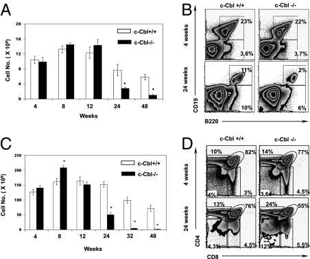
Fig. 1.
Diminished lymphocyte development in older c-Cbl−/− mice. (A) Absolute numbers of CD19+ B cells of the BM were determined as average per animal (two tibia and fibula) at indicated weeks of age. Each group contains n = 5 mice. Data are representative of five independent experiments. Data are presented as mean ± SEM. *, P values <0.05. (B) FACS plots indicating relative frequencies of B cells at 4 (Upper) and 24 weeks (Lower) of age. Data are representative of 10 independent experiments. (C) Absolute numbers of thymocytes at indicated weeks of age. Each group contains n = 5 mice. Data are representative of 10 independent experiments. Data are presented as mean ± SEM. *, P values <0.05. (D) Analysis of thymic T cell development at 4 (Upper) and 24 weeks of age (Lower) in c-Cbl−/− mice.

