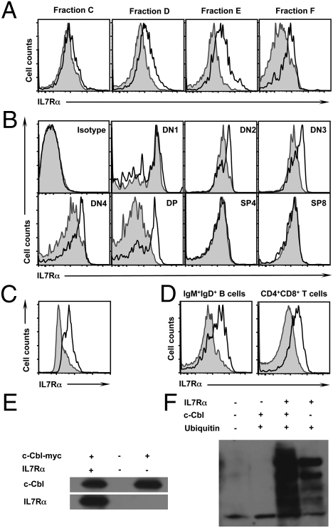
Fig. 5.
Defective IL7R down-regulation in c-Cbl−/− cells. (A and B) Surface expression of IL7R in Hardy Fractions of the BM (A) and T cell subsets of the thymus (B) of 24-week-old mice. Filled histograms represent c-Cbl+/+ cells and open histograms represent c-Cbl−/− cells. (C) Surface expression of IL7R in in vitro differentiated B cells from HSCs. Filled histograms represent c-Cbl+/+ cells and open histograms represent c-Cbl−/− cells. (D) Surface expression of IL7R in pregated B220+CD43−IgM+IgD+ (Fraction F) cells of the BM (Left) and CD4+CD8+ (DP) cells of the thymus (Right) of 24-week-old mice. Filled histograms represent c-Cbl+/+ cells and open histograms represent c-CblA/− cells. (E) Coimmunoprecipitation studies. 293T cells were cotransfected with plasmids expressing c-Cbl and IL7R proteins. Cell lysates were coimmunoprecipitated using myc antibodies. Precipitates were subjected to immunoblotting and detected using c-Cbl (Upper) and IL7R (Lower) antibodies. (F) Ubiquitylation assays. 293T cells were cotransfected with plasmids expressing c-Cbl, IL7R, and ubiquitin proteins. Cell lysates were immunoprecipitated using IL7R antibodies. Precipitates were subjected to immunoblotting and detected using ubiquitin antibodies.

