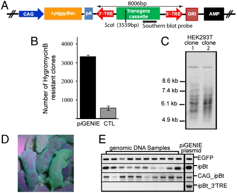
Fig. 1.
piGENIE supports cell transfections and animal transgenesis. (A) Schematic representation of piGENIE. The transposon cassette is delimited by the 3′- and 5′-TREs. Transposon size, restriction sites within the transposon, and Southern blot probe location are indicated. (B) 0.5 × 10⁵ HEK293Tcells were transfected with 400 ng of piGENIE plasmid. As control for random nontranspositional integration of the plasmid HEK293T cells were transfected with 400 ng of a construct (piGENIE/ΔpiggyBac) lacking a functional pBt gene. Transposition activity was measured by counting methylene blue stained, hygromycin-resistant colonies after a three-week selection period. Data are shown as mean values with SD (N = 3). (C) Southern blot analysis of gDNA from a clonal expansion of transfected cells to analyze insertion events. Samples were digested with ScaI and BamHI and probed with a DIG-labeled DNA fragment corresponding to the EGFP gene. (D) Depiction of mosaic piGENIE transgenic mice. (E) Assessment of plasmid backbone insertions into the genome of ten piGENIE mice by PCR analysis (piGENIE plasmid served as a control). The analysis revealed that whereas all animals are transgenic for EGFP (top panel), they also displayed ipBt gene insertions (second panel from the top). Moreover, amplification products for the ipBt gene with its CAG promoter were obtained (second panel from the bottom) but not from a region spanning the ipBt gene and the 3′-TRE (bottom panel), indicating nontranspositional insertion of the plasmid backbone only but not of the entire plasmid.

