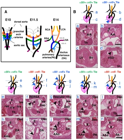
Fig. 3.
Cardiovascular defects in α5/αv-cdKO embryos. (A) Schematic of the embryonic remodeling process of the symmetric branchial arch arteries into the aortic arch and great vessels (E14.5). Some vessels regress (gray dashed lines), while others are stabilized. (Ba-n) Sections and schematics illustrating the heart and great-vessel defects found in integrin conditional double-knockout (cdKO) and hemizygous (cdHemi) mice. Genotypes (in endothelial cells) are indicated and color-coded for this and subsequent figures as follows: green, α5flox/+; αvflox/+; Tie2-Cre (α5/αv-cdHemi); blue, α5flox/−; αvflox/+; Tie2-Cre (α5-cKO; αv-cHemi); red, α5flox/−; αvflox/−; Tie2-Cre (α5/αv-cdKO). Schematics illustrate the observed phenotypes and the blue dashed lines refer to the locations of the corresponding sections. Control hearts and great arteries (a,c,e,h,k,m); atrophic (or interrupted, not shown) aortic arch and ventricular septation defect (VSD) (b,d); interrupted aortic arch and retro-oesophageal right subclavian artery (RERSA) (f,i); vascular ring (g,j); interrupted aortic arch, loss of ascending aorta and RERSA (l,n). (a) Control aortic arch; (b) atrophic aortic arch; (c) separated left and right ventricles; (d) VSD; (e) normal position of great arteries ventral to the trachea; (f) retro-oesophageal right subclavian artery (arrowhead); (g) vascular ring around trachea (arrowhead), which results from maintenance of left sixth branchial arch artery and left dorsal aorta in A; (h) aorta in plane of ductus arteriosus (DA); (i) tiny remnant of ascending aorta (arrowhead) in plane of DA; (j) maintenance of both left and right sixth branchial arch arteries resulting in vascular ring; (k) control ascending aorta in plane of DA; (l) absence of ascending aorta in plane of DA; (m) control ascending aorta in plane of pulmonary artery; (n) remnant of ascending aorta in plane of pulmonary artery (arrowhead). AA, aortic arch; AAo, ascending aorta; B, bronchi; Dao, descending aorta; E, oesophagus; lca, left carotid artery; lsa, left subclavian artery; LV, left ventricle; rca, right carotid artery; rsa, right subclavian artery; RV, right ventricle; T, trachea; Th, thymus. Scale bars: 200 μm.

