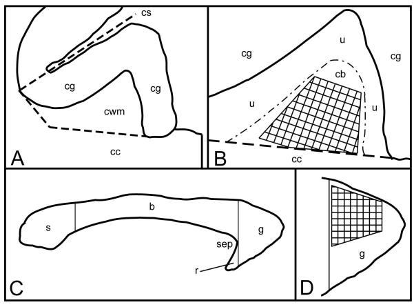
Figure 1.
This figure depicts the cross-sectional areas for the cingulate white matter (A and B) in the coronal plane, and the genu of the corpus callosum (C and D) in the mid-sagittal plane. A: The cingulate gyrus and underlying white matter at the level of the anterior commissure. The cingulate white matter (cwm) is bounded dorsally, medially, and laterally by the cingulate cortex (cg). Ventrally, it is bounded by the body of the corpus callosum (cc). An inferolateral boundary was arbitrarily defined by a line oriented perpendicular to the cingulate sulcus (cs), from the gray matter/white matter boundary dorsally, to the underlying corpus callosum (cc) ventrally. B: Enlarged diagram of the cingulate white matter from A. The cingulate white matter is composed of both the cingulate bundle proper (cb) and local system fibers (u) underlying the cingulate cortex (cg). The hatched area represents the portion of the cingulate bundle sampled for electron microscopic analysis. C: A mid-sagittal diagram of the corpus callosum of the rhesus monkey. The genu (g) is the anterior-most portion of the corpus callosum. Its posterior boundary is defined by a straight line, perpendicular to the long axis of the corpus callosum, at the boundary of the corpus callosum with the anterior tip of the septum pellucidum (sep). Splenium (s), body of the corpus callosum (b), rostrum (r). D: An enlarged diagram of the mid-sagittal area of genu of the corpus callosum in C. The hatched area represents the dorsal portion of the genu sampled for electron microscopic analysis.

