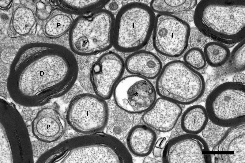
Figure 5.
Electron micrograph from the cingulate bundle of a 31 year-old male rhesus monkey (AM091). Internodal (I) and paranodal (P) profiles of myelinated nerve fibers are indicated. The axon of one myelinated nerve fiber (asterisk) is degenerating. Another myelinated nerve fiber (D) is surrounded by sheath containing dense cytoplasm. Scale bar= 1μm.

