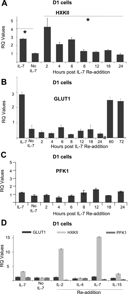
Fig. 4.
Readdition of IL-7 restores gene expression of HXKII. A–C: D1 T cells were deprived of IL-7 for 18 h and washed, and then IL-7 (50 ng/ml) was readded for the periods of time specified in the figures. Total RNA was isolated and transcribed to cDNA as described in materials and methods. qPCR was performed to analyze the gene expression of HXKII (A), GLUT-1 (B), and PFK-1 (C). RQ values were calculated from qPCR data to show relative gene expression as explained in materials and methods. D: D1 T cells were deprived of IL-7 for 18 h then incubated with IL-2, IL-4, IL-7, and IL-15 at an optimal concentration (50 ng/ml) for 2 h. Total RNA was isolated and transcribed to cDNA, and gene expression was analyzed by qPCR as described above. RQ values were calculated from qPCR data to show relative gene expression as explained in materials and methods. The calibrator sample chosen to determine RQ values was the 18-h time point without IL-7. Results are representative of at least 3 independent experiments and values represent average ± SD. *P < 0.05 compared with without IL-7.

