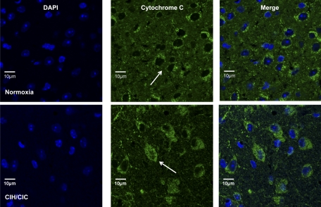
Fig. 3.
P2 CD-1 mice were exposed to intermittent hypoxia/hypercapnia for 10 days, and brain cryosections were stained with an antibody to cytochrome c. Top: coronal sections from mice exposed to normoxia. Bottom: coronal sections from mice exposed to CIH/CIC showing DAPI in blue and cytochrome c in green. Arrows indicate cytochrome c-positive cells.

