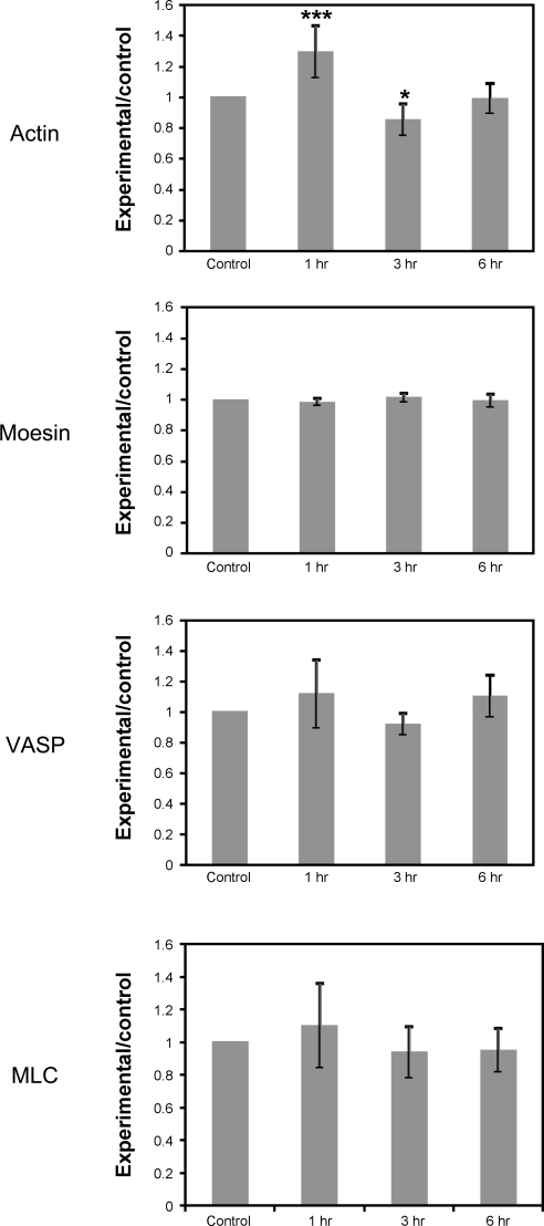
Fig. 1.
iTRAQ proteomics confirmation of protein changes following hypoxic treatment. We found that our proteomic analysis, while identifying a similar set of proteins each time it was performed, was quite variable in the degree of change seen between data sets. Therefore we also used in-cell Western blot (ICWB) analysis to confirm changes seen in our proteomic data sets (data not shown). Actin levels as detected by iTRAQ were slightly elevated after 1 h of hypoxia, then fell at 3 and 6 h. Moesin, vasodilator-stimulated phosphoprotein (VASP), and myosin light chain (MLC) were unchanged in iTRAQ analysis. Data are presented as means ± SE for 3 iTRAQ experiments. A significant change was only found for actin levels after 1 h of hypoxia by one-way analysis of variance. *P < 0.05, ***P < 0.001 vs. control.

