Figure 1.
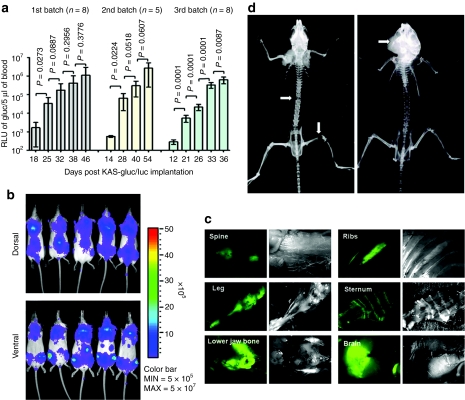
Characterization of the KAS 6/1 disseminated human multiple myeloma SCID mouse model. ICR-SCID mice were injected intravenously via the tail vein with 6 × 10⁶ KAS 6/1-Gluc-CFP cells. (a) Tumor growth in mice from three independent experiments was monitored by measuring Gaussia luciferase (Gluc) activity in 5 µl of whole blood (n = 5–8 mice per batch). P < 0.05 denotes significant increase in tumor burden compared to the previous time point. (b) Bioluminescent imaging for firefly luciferase activity showing distribution of human myeloma disease in bone marrow of mice. (c) Images of explanted tissues showing presence of CFP+ tumors predominantly in the spine, legs (femur), lower jaw bones (mandible). (d) X-ray images taken of the skeleton of mice with advanced myeloma disease indicate presence osteolytic lesions or fractures (white arrows) in the spine, legs, or mandible due to myeloma growth. CFP, cyan fluorescent protein; SCID, severe combined immunodeficiency.

