Figure 2.
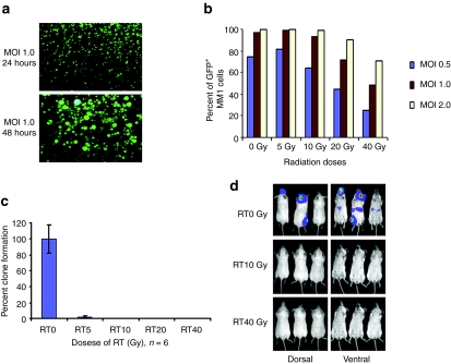
Feasibility of lethally irradiated MM1 cells as a vehicle for MV delivery. (a) 10 Gy lethally irradiated MM1 cells remained susceptible to MV-GFP infection and expressed viral proteins, resulting in syncytia formation 48 hours postinfection. (b) Quantitation of viral infection and expression in irradiated cells by measuring percentage of single cells expressing GFP using flow cytometry. (c) Clonogenic assays showed that irradiation (RT) resulted in significant loss of MM1 cell viability and clonogenicity at 14 days after cell plating (n = 6 replicates per RT condition). RT0, mock irradiated. (d) Bioluminescent images showing tumor growth in mice injected intravenously with nonirradiated (RT0 Gy) MM1-Fluc cells. In contrast, no tumor growth was seen in mice given lethally irradiated (RT10 or RT40 Gy) MM1-Fluc cells at 42 days after cell infusion. Fluc, firefly luciferase; GFP, green fluorescent protein; MM, multiple myeloma; MOI, multiplicities of infection; MV, measles virus.

