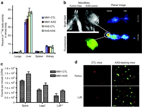
Figure 3.
Biodistribution of MM1 cell carriers after intravenous delivery in SCID mice-bearing disseminated KAS 6/1 myeloma disease. (a) Percentage of 111Indium oxine-labeled MM1 or KAS 6/1 cells in organs harvested from tumor-free SCID mice (CTL) or mice with disseminated KAS 6/1 disease (KAS). (b) Whole body planar gamma camera image of tumor free or tumor-bearing mice given 111Indium oxine-labeled MM1 cells. The hot spot in the gamma camera image (circled) correlated with the presence of a large CFP+ KAS 6/1 tumor in the mandible. (c) Dosimetry measurements (counts per minute) showing the amount of radiolabeled MM1 cells in a portion of the spine, legs, or mandibles of KAS 6/1-bearing mice as compared to normal control (CTL) mice. (d) Fluorescence microscopy confirmed presence of DiI-labeled (red) MM1 cells in the bone marrow flushings obtained from the femurs and lower jaw bones (LBJ) of tumor free and KAS 6/1 (CFP+)-bearing mice. CFP, cyan fluorescent protein; IN, indium; MM, multiple myeloma; SCID, SCID, severe combined immunodeficiency.

