Figure 4.
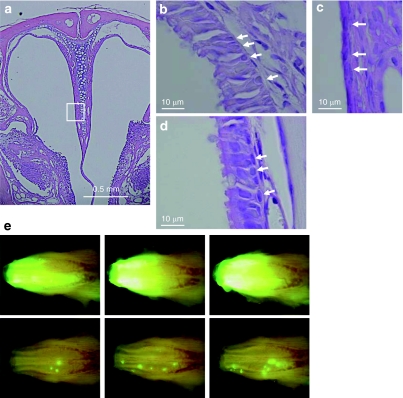
Clustering of transduced cells after the polidocanol-mediated stripping of epithelial cells followed by rapid regeneration. Mouse nasal tissue was perfused with 10 µl of 2% (vol/vol) polidocanol (n = 3). (a) Representative low-power view (original magnification ×50) of the nasal cavity 24 hours after perfusion. Respiratory epithelium, marked by a white box was further magnified (original magnification ×200). The respiratory epithelium before the treatment is shown in b. Arrow indicates basal cells. The respiratory epithelium was completely stripped 24 hours after polidocanol perfusion, whereas the basal cell layer was (c) retained and (d) regenerated 7 days after treatment. (e) This treatment was done after transduction with F/HN-SIV vector. Seven days after transduction of nasal epithelial cells with F/HN-SIV-GFP (4 × 10⁸ transduction units/100 µl/mouse), the nasal epithelium was stripped via perfusion with 10 µl of 2% (vol/vol) polidocanol. Polidocanol treatment was repeated again 3 weeks later. Histological sections were analyzed 58 days after vector administration (30 days after the last polidocanol treatment). In situ imaging of GFP expression in the nasal cavity of untreated mice (top panel in e) or mice treated with polidocanol (bottom panel in e). Clusters of GFP-positive cells were seen in the polidocanol-treated mice. GFP, green fluorescent protein.

