Figure 2.
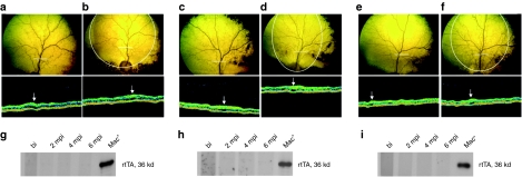
Assessment of retinal morphology and evaluation of humoral immune response against the tTA2 and rtTA.M2 proteins. (a,b) Fundus photography and OCT image of the AAV2/4TetOff.rpe65 treated eye of A2 before (a) and 6 months postinjection (b). (c,d) Fundus photography and OCT image of the AAV2/4TetOn.rpe65 treated eye of A4 before (c) and 6 months postinjection (d). (e,f) Fundus photography and OCT image of the AAV2/4CMV.rpe65 treated eye of A6 before (e) and 6 months postinjection (f). The white circle on the fundus photography indicates the zone of the bleb. The white line on the fundus photography indicates the OCT scanning path. The white arrow on the OCT image indicates the retinal vessel. (g–i) Evaluation of antibody generation against tTA2 and rtTA.M2 before and at 2, 4, and 6 months postinjection in A2 (g), A4 (h), and A6 (i). bi, before injection; mpi, months postinjection; OCT, optical coherence tomography.

